
Something like this.
Right side forehead smooth, eye pulled down - full of tears.
Keratinized and angry looking conjunctiva, man complained of 'hard of hearing' and loss of light perception and sensation from his right eye.
The man smelled certain way and I noticed the old stained overall holding his pants.
After he was seen, instead of ectropion repair, Medial Canthal Tendon repair was to be go ahead with it.
I've never seen the procedure and normally this would be done under Major OR (anesthetic)- but since this man had lost all sensation in his right face - including in his cornea - it was ok.
During the surgery - Dr.Johnson says,
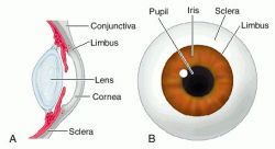
"the one thing that I learned after medical school was that corneal cell regenerates as there are stem cells in limbus - so this man- not being able to feel anything indicates that this is not a normal bell's palsy - It's not just the CN 7 that's affected."
and then he asks me:
"which CN is the one for corneal sensation?"
of course I go, "um..."
"CN 5, trigeminal nerve" this was new to me. I think back from my book if I had ever read something about this. Nope, I didn't see it (later that night I went back to my text book and of course it doesn't say anything... through google I just confirmed this - as I almost wrote CN3 - oculomotor nerve)
And then the man says, "my hearing is bad in one ear (indicating his right where the bell's palsy was)"
Dr.J exchange looks with me and says, "and also the CN 8 - there's something going on."
I noticed that near his jaw and neck, there's a overly protruding bone (?) and I point at that as I wonder.
Dr.J nods then says "five."
No light perception..I wonder if his right pupil constricted? because this would indicate that his CN3 was also affected.
Back to limbal epithelial cells of the cornea.
During undergrad, I had the most difficulty wrapping around the concept of stem cells and to this day, I'm quite confident to say I don't understand whole lot what's going on with the cell development.
I know that there are different levels of stem cells such as totipotent (total: can make an individual!!) and pluripotent (sublevel: not fixed as to developmental potentialities; especially : capable of differentiating into one of many cell types) cells and then it gets complicated....
To read the "Limbal epithelial stem cells of the cornea" click here!
Summary for myself (short copy and paste from the above link):
Ok, so what is cornea?
The cornea is responsible for protecting the eye against insults such as injury and infection. It also provides the majority (two thirds) of the total refractive power of the eye and is therefore the major refracting lens (Meek et al., 2003). So the LASIK, LASEK surgery is basically carving the corneal to correct the refractive power.
The corneal epithelium is a dynamic physical barrier preventing the entry of deleterious agents into the intraocular space. It consists of superficial squamous cells, central suprabasal cells and a single layer of inner columnar basal cells (Seckera& Daniels 2009).
Corneal integrity and therefore function is dependent upon the self-renewing properties of the corneal epithelium. The prevailing hypothesis is that this renewal relies on a small population of putative stem cells located in the basal region of the limbus (Seckera& Daniels 2009).
that's what Dr.J said!!!
Throughout life, our self-renewing tissues rely upon populations of stem cells / progenitors to replenish themselves throughout life following normal wear and tear and injury. The corneal epithelium on the front surface of the eye is no exception as dead squamous cells are constantly sloughed from the corneal epithelium during blinking. At the corneo-scleral junction in an area known as the limbus, there is a population of limbal epithelial stem cells (LESCs). LESCs share common features with other adult somatic stem cells including small size (Romano et al., 2003) and high nuclear to cytoplasmic ratio (Barrandon and Green, 1987). They also lack expression of differentiation markers such as cytokeratins 3 and 12 (Kurpakus et al., 1990; Schermer et al., 1986).
No comments:
Post a Comment